Neurodiagnostics and Neuroradiology
From imaging tests to EEGs, our doctors use many tests to understand neurological disorders.
At Memorial Neuroscience Institute, our neurologists and neurosurgeons use imaging and other tests to better understand your symptoms and determine what’s causing them. Neurological conditions can affect everything you do, so getting the right care can provide relief and help you get back to the things you enjoy most. That process starts with an accurate diagnosis.
Diagnosing Neurological Conditions
When you come to us for care, you’ll find a team approach that may include experienced doctors including Neuroradiologists- Imaging specialists with expertise in conditions affecting the brain and spine; and Pathologists- Specialists who analyze tissue samples, who know which tests to order and work together to interpret the results.
Our neuroradiologists use leading-edge equipment to provide clear pictures. We individualize scans to each patient's unique medical history and symptoms and strive to provide a diagnosis as quickly as possible so you can start treatment.
 American College of Radiology (ACR) accreditation
Memorial Healthcare System maintains ACR accreditation in CT, MRI and other imaging tools. These efforts reflect our commitment to meeting the highest standards in safety, quality and staff qualifications.
Using imaging tests that help us view the structures inside your body, our neuroradiologists look for problems in and around the brain and spine, including:
- Damage to bones, nerves and soft tissues
- Issues with blood vessels and blood flow
- Tumors, cysts and other lesions
Specialized neuroimaging tests for diagnostic and treatment purposes, including:
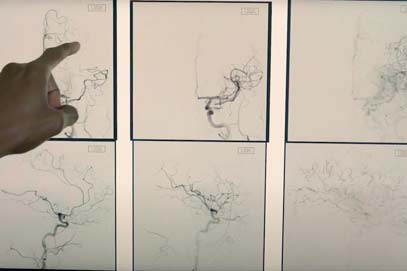
CT and MR angiography help doctors evaluate your blood vessels. They combine traditional CT or MRI imaging with an injection of contrast dye into a vein. The dye provides a better view of the blood vessels and any irregularities.
These tests show how well blood is flowing into brain tissues. They require the precisely timed injection of contrast dye into a vein, followed by a CT or MRI scan.
This MRI technique evaluates nerve pathways and connections in the brain. It helps doctors identify impaired pathways, such as in multiple sclerosis (MS) and stroke, and measure disease progression. Neurosurgeons also use diffusion tensor imaging to map critical brain areas before surgery.
Functional MRI measures changes in blood flow when your brain is active. This test helps assess the effects of diseases, such as trauma, stroke and Alzheimer’s disease, on your brain. It’s also used in surgical planning to map brain areas responsible for critical functions.
This specialized MRI measures chemical changes in the brain, especially those caused by brain tumors, skull base tumors and epilepsy.
Myelography helps doctors identify problems with your spinal cord and the nerve roots, such as herniated discs and other causes of back and neck pain. This test involves injecting a contrast material into the space around the spinal cord and then taking specialized X-ray images.
Discography is a procedure to find the source of back or neck pain. Your doctor injects a contrast liquid into a suspect disc and then takes an X-ray or CT scan. If the injection reproduces symptoms, it suggests the disc is the source.
Other Neurological Tests
Additional imaging studies we use include conventional X-rays as well as CT, MRI and PET scans. Other tests we use to diagnose neurological conditions include:A neurological exam is a group of tests to find out how well your brain and nervous system are working. These tests look at:
- Involuntary processes, such as breathing, heart rate and digestion
- Muscle reflexes, balance and coordination
- Senses, such as touch, hearing, smell and vision
- Thinking and memory
An EMG test helps detect muscle damage. It is performed by inserting a fine needle into a muscle to measure electrical activity. This test is usually performed with a Nerve Conduction Study to measure how electrical signals move through your muscles and nerves,
EMG and nerve conduction studies are common tests to diagnose and determine the severity of certain neurological conditions such as:
A Nerve conduction study involves placing electrodes on your skin at various places around your body. One set of electrodes generates a mild pulse of electrical activity. The other measures a muscle’s response. Nerve conduction is the time between the pulse and response. Low conduction is a sign of nerve damage. This test is usually performed with an EMG to measure how electrical signals move through your muscles and nerves.
EMG and nerve conduction studies are common tests to diagnose and determine the severity of certain neurological conditions such as:
An EEG is a painless, noninvasive test to measure brain electrical activity. It’s an essential test for diagnosing epilepsy.
In this test, we place small metal discs (electrodes) on your scalp to detect electrical activity. Thin wires attached to the discs carry the information to a computer monitoring system. You may have an EEG in your doctor’s office or in our epilepsy monitoring unit.
Stereotactic EEG is a minimally invasive surgical procedure for people with drug-resistant epilepsy. It provides more detailed information about seizure activity and can help determine if you might benefit from surgical epilepsy treatments.
The neurosurgeon places thin wire electrodes deep in your brain through tiny holes in your skull. We use image guidance and a robotic arm to insert the electrodes precisely. After we place the electrodes, you spend time in our epilepsy monitoring unit to record your brain activity.
This noninvasive test measures the transmission of nerve signals to your brain. Electrodes placed on the skin over your muscles deliver small electrical pulses. Electrodes on your scalp record the pulses as they reach your brain.
Evoked potentials tests can detect slow transmission of electrical signals due to nerve damage, such as in MS and other neuroimmune conditions.
A lumbar puncture is a procedure to collect a sample of the fluid from around your spinal cord. Your provider inserts a fine needle between the two lower spinal bones (vertebrae) to obtain the sample and send it to a laboratory for analysis. This test is typically used to check for signs of cancer, infection and inflammation.
You may have a lumbar puncture in your doctor’s office. If you’re in the hospital, our neuroradiologists perform the procedure using X-ray imaging to guide needle placement.
A biopsy is a procedure to collect a tissue sample for laboratory analysis. We use biopsies to diagnose and monitor:
- Neuromuscular disorders: Our doctors perform skin, muscle and nerve biopsies for these conditions. A skin biopsy involves collecting a small skin sample to measure the density of nerve fibers. Muscle and nerve biopsies are performed surgically. Pathologists analyze these tissues for signs of damage, disease or change in function.
- Brain and spine tumors: These biopsies usually require surgery. Whenever possible, the surgeon removes as much of the tumor as possible to avoid a second surgery. For some spine tumors, we may be able to perform a needle biopsy. This procedure involves inserting a fine needle into the tumor to remove a tissue sample.
We use a wide range of laboratory tests when evaluating neurological disorders. These tests may help us identify an underlying cause of a neurological condition, such as diabetes, hormone changes and vitamin deficiency. Blood tests are also available to measure neurological disease activity, including inflammation, cancer and autoimmunity.
Genetic testing can detect specific changes in the DNA of brain, skull base and spine tumor cells. Doctors use these tests to better understand tumors and select the most effective treatments.
Neuropsychological evaluations involve a series of tests that measure different aspects of brain function. Our neuropsychologists select and administer these tests and interpret the results. Testing can help diagnose certain neurological conditions and track changes in how your brain functions.
Neurodiagnostic Testing: Why Choose Memorial Neuroscience Institute?
Neurological testing is part of the exceptional care you’ll find at Memorial Neuroscience Institute, featuring:
- Skilled specialists: Your team includes fellowship-trained neurologists, neurosurgeons and neuroradiologists. They use their extensive expertise and experience to diagnose and treat neurological disorders.
- Leading-edge tests: We leave no stone unturned to find the source of your condition. That may include a sophisticated procedure or advanced imaging test using a multislice CT scanner or 3T MRI.
- Personalized approach: Our doctors tailor testing to your medical needs. We offer both conventional and highly specialized tests to guide your care.
Making a Difference for Our Patients
Related Providers
-


 Norman Ajiboye, MD Neuro-Interventional, Vascular Interventional Neurology
 -
 Brandon Davis, MD, PHD Neurosurgery, Neuro-Interventional
 -
 Hyun Woo Kim, MD Neuro-Interventional, Vascular Interventional Neurology